This Article
Citations

Except where otherwise noted, this work is licensed under Creative Commons Attribution-NonCommercial 4.0 International License.
Interesting Images 3
Keywords: Tetralogy Fallot ; Pulmonary Atresia
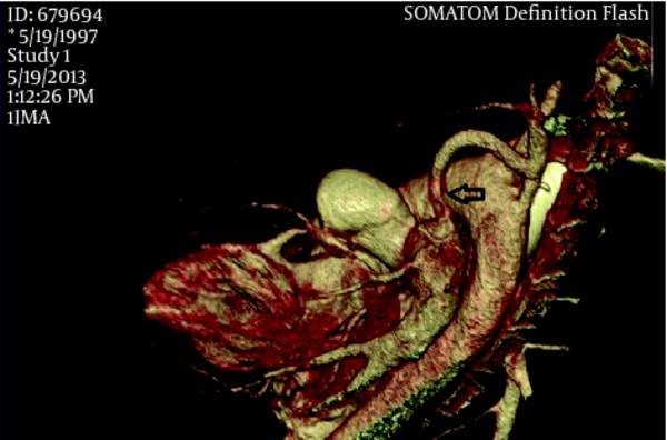
A 16 year old boy that was found to be cyanosed after birth and diagnosed with Tetralogy fallot and pulmonary atresia referred to our adult congenital heart disease clinic with a history of palliative surgery in childhood and recent dyspnea on exertion, New York Heart Association (NYHA) functional class III. He was cyanosed with marked clubbing of the fingers and toes. Oxygen saturation in room air was %64.
Based on the Cardiac CT image what is the diagnosis?
|
Figure 1
Echocardiography Images
|
Answer: Occluded left BT shunt
Image reconstruction by volume rendering and editing software create 3D images with removed overlying structure that adequately visualize structure of interest. The 3D reconstructed image seems attractive regarding the ease of interpretation but it is essential to know the limitation of reconstruction technique and it is important to continually refer back to the 2D axial data set before making a diagnosis(1).
Footnotes
References
- 1. Budoff M, , Shinbane J. Handbook of cardiovascular CT: Essentials for clinical practice Springer; 2008.
 Home
Home Archive
 Search
 Sign In
 Site Menu
 Email this article to a friend


