 Home
Home Archive
 Search
 Sign In
 Site Menu


This Article

Citations
 CrossRef (0)
 - Google Scholar
-
Scopus (0)

Except where otherwise noted, this work is licensed under Creative Commons Attribution-NonCommercial 4.0 International License.
Simultaneous Left Ventricle and Left Atrial Appendage Thrombi in a Patient with Dilated Cardiomyopathy
Keywords: Dilated Cardiomyopathy; Echocardiography; left Ventricle; Thrombus
1. Introduction
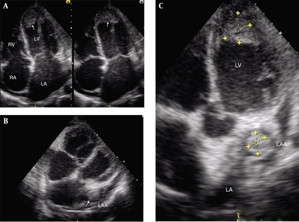
A 24-year-old male, diagnosed with post myocarditis dilated cardiomyopathy (DCM), presented with dyspnea and body swelling. On examination, he was ill-looking. Blood pressure was 90/60 mm Hg and his heart rate was 110 beats per minutes. Upon physical examination, pitting edema was noted in bilateral lower extremities and a S3 gallop sound was audible. Jugular venous pulse was raised. Chest X-ray showed cardiomegaly with pulmonary congestion. Electrocardiography showed sinus tachycardia. The cardiac enzymes were within the normal range. What is the echocardiographic diagnosis based on the Figure 1 and videos 1,2?

|
Figure 1.
Transthoracic Echocardiography
|
2. Answer
Transthoracic echocardiography revealed global wall hypokinesia, dilated chambers, and the left ventricular (LV) systolic dysfunction with moderate pericardial effusion. There was a large thrombus in the LV apex (Figure 1 A, Video 1). In the modified subcostal view, there was another thrombus floating in the left atrial appendage (LAA) (Figure 1 B, 1C, Video 2).
Transthoracic Echocardiography: Apical 4-Chamber View Showing Thrombus in the Left Ventricular Apex
Transthoracic Echocardiography: Modified Subcostal View Showing Thrombus in the Left Atrial Appendage
3. Comments
Patients with DCM are at a particularly high risk of the LV thrombus formation because of the poorly contracting ventricle that allows blood stasis. Atrial fibrillation (AF) is known to be highly associated with the LAA thrombus formation. However, simultaneous thrombus in the LV and LAA is an unusual occurrence in the absence of AF. As LAA is larger, it may be an additional source of thrombi in patients with DCM (1). It has been reported that the LAA maximal area is an independent predictor of LAA thrombi in patients with DCM in sinus rhythm (2). Because of the high prevalence of embolization, anticoagulant therapy should be started immediately. Despite reports of successful resolution with anticoagulants treatment, some experts suggest surgical removal of the thrombus with simultaneous treatment of the underlying cause is the treatment of choice because anticoagulation alone can result in fragmentation of the thrombus and embolization (3).
Footnotes
References
- 1. Bakalli A, Kamberi L, Pllana E, Zahiti B, Dragusha G, Brovina A. The influence of left ventricular diameter on left atrial appendage size and thrombus formation in patients with dilated cardiomyopathy. Turk Kardiyol Dern Ars. 2010;38(2):90-4. [PubMed]
- 2. Bakalli A, Georgievska-Ismail L, Kocinaj D, Musliu N, Zahiti B, Krasniqi A, et al. Left ventricular and left atrial thrombi in sinus rhythm patients with dilated ischemic cardiomyopathy. Med Arch. 2012;66(3):155-8. [PubMed]
- 3. Balaji R, Srinath VS, Krishna Kumar KV. Left atrial ball valve thrombus. J Clin Sci Res. 2013;2:236-8.
")


