This Article
Citations

Except where otherwise noted, this work is licensed under Creative Commons Attribution-NonCommercial 4.0 International License.
Bubbles in Pericardial Fluid: Multimodality Imaging in Iatrogenic Hydropneumopericardium
Abstract
Introduction: The term hydropneumopericardium describes the simultaneous accumulation of fluid and gas in the pericardial sac. This condition is mostly caused by primary infiltrative lesions from the adjacent organs, pericardial infections, or trauma and is a very rare situation, usually with favorable outcomes.
Case Presentation: We describe a female patient with Lutembacher’s syndrome complicated by cardiac tamponade. After surgical treatment, she developed iatrogenic hydropneumopericardium, which was treated conservatively.
Conclusions: Iatrogenic hydropneumopericardium can be managed conservatively with supportive measures, and most of these cases resolve spontaneously if they are not large and destabilizing.
Keywords: Pericardial Effusion; Pneumopericardium; Lutembacher's Syndrome
1. Introduction
Simultaneous accumulation of serous fluid and gas in the pericardial sac is called hydropneumopericardium. This very uncommon condition in adults (1) is mostly a consequence of primary infiltrative lesions from the adjacent organs, pericardial infections, or trauma (2) and usually harbors favorable outcomes. Still, the condition may become severe occasionally (1). Previously, most cases in adults were derived from gas-forming bacterial infections, also known as "pyopneumopericardium". With the advent of wide-spectrum antibiotics, however, this condition has become rare and other unusual causes such as paraesophageal hernia and surgical complications have become more prevalent (2). Here, we report a case of iatrogenic hydropneumopericardium in a patient with Lutembacher’s syndrome presenting with cardiac tamponade.
2. Case Presentation
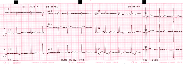
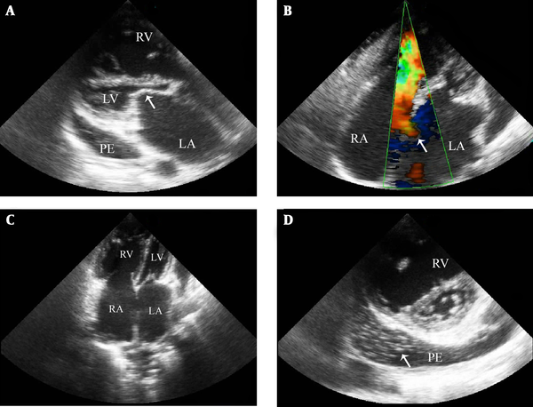
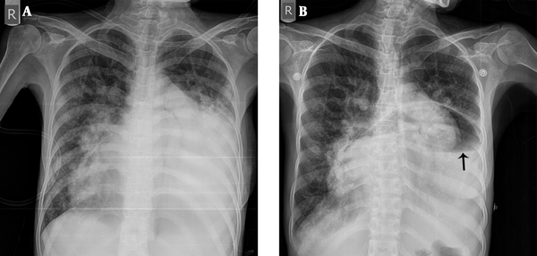
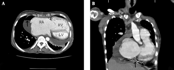
A 55-year-old-woman, a known case of rheumatic mitral valve stenosis and congenital atrial septal defect (Lutembacher’s syndrome), referred to the emergency department complaining of dyspnea at rest. On primary evaluation, she had a blood pressure of 110/70 mmHg with a 15-mmHg paradoxical pulse. The jugular venous pressure was higher than 12 mm of H2O, and lung examination revealed pulmonary rales up to 2/3 of the lung. She was on Digoxin and Propranolol. Her electrocardiogram showed an atrial fibrillation rhythm with a controlled rate (90 beats per minute) alongside signs of right ventricular (RV) hypertrophy, which could be indicative of pulmonary hypertension (Figure 1). Primary echocardiogram showed the presence of rheumatic mitral valve stenosis (Figure 2A), accompanied by a large atrial septal defect (Figure 2B), leading to severe pulmonary hypertension (estimated systolic pulmonary arterial pressure = 100 mmHg) and enlarged dysfunctional RV and right atrium (RA) (Figure 2C), while the left ventricular (LV) function was preserved (ejection fraction = 55%). Also, a massive amount of pericardial effusion was noticed. As the presence of cardiac tamponade was suspected, the patient was sent for the surgical drainage of the pericardial fluid. After the removal of the drainage tubes, follow-up echocardiography revealed the presence of moderate amounts of fluid with a turbulent flow of air bubbles within the fluid (Figure 2D). A new chest radiograph, in comparison with the previous one, showed the appearance of an air-fluid level within the pericardial sac (Figure 3A, 3B). This finding, together with noticeable air bubbles, was also observed in the films from cardiac computed tomography (Figure 4A, 4B). The patient did not complain of dyspnea exacerbation after developing this problem, and the clinical signs of tamponade were absent. The acquired hydropneumopericardium was treated conservatively. A week later, no air was noticed in the pericardial sac on the follow-up echocardiogram.
|
Figure 1.
Electrocardiogram of the Patient, Showing an Atrial Fibrillation Rhythm with a Controlled Rate (90 Beats per Minute) alongside Signs of Right Ventricular Hypertrophy and Pulmonary Hypertension (Right Axis Deviation with qR Pattern in Lead V1, Secondary ST-T Change, and Deep S Waves in Leads V5 and V6)
|
|
Figure 2.
Findings of the Echocardiographic Examination of the Patient
|
|
Figure 3.
Radiologic Findings of the Patient
|
|
Figure 4.
Computed Tomographic Findings of the Patient
|
3. Discussion
Hydropneumopericardium is a very rare condition with a much lower frequency than pneumopericardium or pericardial effusion. Reported causes for this condition are as follows:
1) Most cases of hydropneumopericardium in the past were due to the involvement of the pericardium by gas-forming infectious agents, a process also called "pyopneumopericardium". With the advent of broad-spectrum antibiotics, however, other situations now constitute the more prevalent causes of this disease.
2) Currently, most cases are iatrogenic and secondary to the treatment of pericardial effusion via surgical or catheter-based fluid drainage approaches (1, 3).
3) Open or closed chest wall traumas can lead to the entrance of air into an inflamed pericardium (4).
4) Pneumomediastinum can progress secondary to alveolar rupture as a consequence of increased alveolar pressure (asthma, mechanical ventilation, cocaine inhalation, cough, or intense exercise) into the pericardial sac (5).
5) Drainage may occur from the adjacent hollow viscous organs such as the esophagus, stomach, and bronchial tree secondary to perforation and fistula formation as a complication of neoplasms, peptic ulcer, or even paraesophageal hernia (6).
6) rarely hydatid cysts can infect pericardium (7).
7) Idiopathic conditions have also been reported (2).
In 1931, Shackelford proposed the following criteria for the diagnosis of hydropneumopericardium: A) high-pitched tympanic percussion sound; B) loud metallic splashing sound synchronous with heart sounds; and C) characteristic chest X-ray with an air-fluid level in the pericardial cavity (4). Computed tomographic scan is also a useful tool as a guide to both diagnosis and determination of the etiology. Echocardiography may be able to show the presence of air-fluid interface in the pericardial sac (2); and as was the case in our patient, the presence of air bubbles within the pericardial fluid may also be deemed an echocardiographic sign for the detection of hydropneumopericardium. The course of the disease varies depending on the etiologic cause. Whereas most cases of pyopneumopericardium may experience a malignant and toxic course, the majority of the cases with iatrogenic hydropneumopericardium in the absence of a large and destabilizing hydropneumopericardium tend to resolve spontaneously with supportive measures (2), similar to our case.
Acknowledgments
The authors would like to thank Dr. Nasrin Shokrpour at the Center for the Development of Clinical Research of Nemazee Hospital for editorial assistance.
Footnotes
References
- 1. Peters F, Patel A, Essop R. Iatrogenic hydropneumopericardium. Cardiovasc J Afr. 2012;23(3):e1-2. [DOI] [PubMed]
- 2. Wu M, He X, Yang G. Spontaneous tension hydropneumopericardium complicating serofibrinous pericarditis. Eur J Cardiothorac Surg. 2006;29(3):422-4. [DOI] [PubMed]
- 3. Aidala E, Valori A, Abbruzzese PA. Postoperative hydropneumopericardium. Heart. 2004;90(9):1058. [DOI] [PubMed]
- 4. Rakotoarivelo RA, Razafimahefa SH, Ramanalimanana P, Ranoharison D. , Johnson AD. Rakotoarimanana S. Large uncomplicated hydropneumopericardium. SA J of Radiol. 2010;14(1):19.
- 5. McEachern RC, Patel RG. Pneumopericardium associated with face-mask continuous positive airway pressure. Chest. 1997;112(5):1441-3. [PubMed]
- 6. Grandhi TM, Rawlings D, Morran CG. Gastropericardial fistula: a case report and review of literature. Emerg Med J. 2004;21(5):644-5. [DOI] [PubMed]
- 7. Khosravi Maharlooei M, Attar A, Goran A, Amuee S, Dehghan A, Monabati A. Hydatid Cyst of Ovary: A Case Report Iran J Med Sci. 2009;34(1):76-9.
 Home
Home Archive
 Search
 Sign In
 Site Menu
 Email this article to a friend


